Happy Week 10! I hope everyone had a fun Fourth of July! Midterms are once again upon us as another 5 weeks have blown by. This week, I have a heavy radiology based week, as I have a lecture and lab exam for radiological positioning, radiological critical thinking and orthopedic and musculoskeletal imaging. I thought to keep on theme with these exams, I would talk about one of our practical, hands on imaging classes, radiological positioning and how vital it is to our future practice as chiropractic physicians.
Why Are X-Rays Important for Chiropractic Medicine?
X-rays are valuable images of different aspects of anatomy that can aid in both our diagnosis and treatments of patients. Therefore, it is crucial to conduct excellent image studies, and be able to not only setup the X-ray machine and equipment, but also instruct and position the patient so our image includes all desired structures with adequate quality.
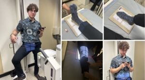
There are many factors, such as FFD, or focal film distance, which describes how far from the X-ray tube is from the Bucky, which helps direct the image to the image detector. If this all seems like technical jargon, it is! But in reality, quality x-rays can save lives, so we must understand them well. In our radiology class, it is organized by body part, or groupings, as we generally take a series of views for each anatomical region.
I really enjoy this class, despite its late day scheduling, due to its practical application and hands on approach. Although tough, radiology is an amazing subject area that we could all benefit from honing.
Read more about Dylan’s Doctor of Chiropractic Medicine student journey.